Knee Injuries

Knee Injuries
By Dr. Virayut Chaopricha
Orthopedic Surgeon at Vibhavadi Hospital
In the year 2004, there was a question raised by those in the golfing community about what happened to Tiger Woods, who had emerged victorious in the Majors from 1999 to 2002, securing seven titles, including four consecutive wins known as the Tiger Slam. However, he had been unable to win any tournaments for a whole year.
Tiger Woods had made a change in his coach at the beginning of 2003, transitioning from Butch Harmon to a new coach, Hank Haney, which sparked much speculation about whether he would have another grand comeback. This change was motivated, in part, by the fact that Tiger Woods had experienced discomfort in his left knee and back due to his previous swing technique. After undergoing arthroscopic surgery in 2002, it was discovered that there was fluid in his knee joint and a tearing of the anterior cruciate ligament (ACL). The ACL plays a crucial role in maintaining the stability of the knee and its mobility during sports activities.
In his book "How I Play Golf," Tiger Woods mentioned a technique he employed to increase his distance by 20 yards. He would snap his left leg straight to achieve a more aligned position, and during the swing, he would generate power by rotating his hips quickly and rotating his left leg while keeping it flexed and then snapping it straight immediately during the downswing. This created a significant torque on his left knee. In 2002, Tiger Woods revealed in an interview with Golf Digest magazine that he experienced knee pain and had to take pain medication before playing. He also mentioned the need to minimize excessive pressure on his knee during the downswing.
It is important to understand the anatomy of the knee joint, its stability, and the management of the muscles surrounding the knee to strengthen and stretch them effectively, reducing the risk of injury.
Anatomy of the knee joint:
The knee joint consists of three bones: the femur (thigh bone), the tibia (shinbone), and the patella (kneecap). The contact surfaces between these three bones are covered by articular cartilage and enclosed within a synovial membrane.
Between the articular surfaces of the femur and tibia, there are C-shaped cushions on the outer and inner sides, known as menisci. They help reduce the impact on the knee joint and contribute to its stability, as well as improving the lubrication of the joint surfaces.
The stability of the knee joint depends on the intactness of the femur and tibia, which form the knee joint, the menisci as cushions, and the surrounding muscles.
The important muscles involved are the quadriceps muscles, located at the front of the thigh, responsible for knee extension, and the hamstring muscles, located at the back of the thigh, responsible for knee flexion. If any of these muscle groups are compromised or not functioning properly, the stability of the knee joint can be compromised.
The stability of the knee joint relies on four major ligaments:
- Lateral collateral ligament: prevents the knee from buckling or tilting sideways.
- Medial collateral ligament: prevents the knee from buckling or tilting inwards.
- Anterior cruciate ligament (ACL): prevents the knee from buckling or sliding forward.
- Posterior cruciate ligament (PCL): prevents the knee from buckling or sliding backward.
The lateral and medial collateral ligaments prevent varus and valgus stress on the knee joint, respectively. The anterior cruciate ligament prevents anterior displacement, while the posterior cruciate ligament prevents posterior displacement of the knee joint.
Perilous conditions that can cause severe knee dislocation or excessive knee movement beyond normal include falls, dislocation, excessive hyperextension or lateral movement of the knee. These incidents can result in a complete tear of the ligaments that hold the knee joint, leading to a single or multiple ligament tears or injuries along with fractures of the joint surface. Additional injuries such as a dislocated knee pillow or supporting cushion can also occur.
The severity of ligament tears can be categorized into three levels: Level 1 involves tearing within the tissue of the ligament, but the ligament is still intact or the tear is not clearly visible. Level 2 indicates partial tearing of the ligament. Level 3 signifies complete separation of the ligament.
Symptoms and findings: In general, knee injuries from playing golf are usually not severe. Apart from falling or stumbling, such injuries are often associated with standing and striking the ball on uneven terrain. It is common to find injuries in the left knee of right-handed golfers due to the significant torsion and weight-bearing on the left knee during swings.
Severe injuries are more frequently encountered in sports such as football, rugby, and tennis. The severity of symptoms may vary depending on the extent of the injury, such as:
- Swelling and pain around the knee joint
- Tender pain at the site of ligament tear, along the joint line, which is the position of the knee pillow
- Inability to bear weight or walking with significant abnormal pain
- Restricted knee flexion or extension, with the knee getting stuck in certain positions, potentially due to a missing knee pillow obstructing the joint
- Sensation of swelling or deformity in the knee, which may result from a complete ligament tear or fractured bones
Medical examination findings:
- Examination of joint fluid or blood within the knee joint, which often presents significant swelling
- Evaluation of the stability on the lateral sides of the knee joint
- Assessment of anterior-posterior stability of the knee joint
- Assessment of range of motion, knee rotation, to determine if the knee pillow is missing or not
Image 3: Evaluation of anterior-posterior stability of the knee joint in the supine position with the knee flexed at 90 degrees. If pulling the tibia forward results in anterior displacement, it indicates an anterior ligament tear. If pushing the tibia downward leads to posterior displacement, it indicates a posterior ligament tear.
Knee X-ray: Performed to identify any fractures or joint dislocation.
Arthroscopic surgery: It is a minimally invasive procedure used to examine the interior of the knee joint, allowing for an accurate and clear diagnosis. Most patients receive an injection into the posterior capsule to numb the lower part of the body, enabling them to undergo surgery without feeling pain. A tourniquet is applied to the upper leg to prevent bleeding during the surgical procedure.
During the surgery, a hole is drilled near the front of the knee, close to the patellar tendon, and a 4.5-millimeter metal tube with lenses and fiber optics is inserted into the knee joint. This allows for visualization of the internal structures of the knee joint, which are displayed on a television screen. The magnification can range from 5 to 10 times the actual size, and the procedure can be recorded as a video for future reference.
Image 4: Arthroscopic Surgery for Knee
Abnormalities Examination:
When examining the knee through arthroscopic surgery, various conditions can be observed, such as soft cartilage, front ligament, back ligament, knee pad, and joint tissues, in order to identify abnormalities or irregularities. These conditions can be corrected through surgical intervention, such as smoothing the cartilage and removing the right knee pad, front ligament, and back ligament.
Postoperative Care:
After the wound has healed well, the patient's knee will be wrapped with thick bandages and compressed with an elastic bandage to reduce swelling for approximately 3-4 days. Then, the patient can begin to exercise and strengthen the quadriceps muscles.
Muscle Conditioning and Physical Therapy after Surgery
- Once the wound has healed properly, the patient can start exercising to strengthen the quadriceps muscles.
- Start muscle conditioning without putting weight on the knee, such as swimming or cycling.
- Muscle conditioning using weights (weight training).
Muscle Stretching Exercises Quadriceps Stretching:
Stand near a wall or table, bend one knee, and bring the foot towards the back, feeling a stretch in the front part of the extended leg. Hold this position for 5-7 seconds, repeating 6-10 times on each side.
Hamstrings Stretching:
Practice bending one knee and extending the other leg, leaning forward and backward to feel a stretch in the back of the extended leg. Hold this position for 5-7 seconds, repeating 6-10 times on each side.
Muscle Strengthening Exercises Quadriceps Muscles:
Sit on a high chair, bend the knee, lift the foot, and extend the knee against resistance. Sandbags or resistance bands may be used.
Hamstrings Muscles: Lie face down, bend the knee, and resist against the force. Weights can be tied to the ankles or resistance can be provided using resistance bands.
Recommendations for Golfers:
-
- Warm up and stretch the muscles to increase flexibility and joint mobility before playing golf.
- Start exercising slowly and gradually increase intensity.
- Avoid overexertion or causing injury during exercise.
- Consistently work on strengthening the muscles.
- Play golf within your own limits, considering time, tempo, and balance as important factors. This will greatly reduce the risk of injury.
- If you experience knee pain or abnormal movement, seek medical attention for proper diagnosis and treatment.
Related Packages

Package รักษาโรคกระดูกสันหลังด้วยวิธีเวอร์ติโบรว์พลาสตี้ (Vertebroplasty)
The treatment for vertebral compression fractures caused by osteoporosis using Vertebroplasty involves injecting bone cement into the fractured vertebrae. Vibhavadi Hospital has created a Vertebroplasty treatment package as an option for treating this condition.

Package ฉีดน้ำหล่อลื่นผิวข้อเข่า Artificial Synovial Fluid Injection
The treatment of osteoarthritis has advanced significantly in recent years. In cases where oral medication and physical therapy do not yield satisfactory results, intra-articular injection is an alternative option. Vibhavadi Hospital offers a Knee Joint Lubrication Injection Package, which helps improve joint lubrication and reduce friction. Each injection is administered one week apart, providing effects that last for 9 months to 1 year.
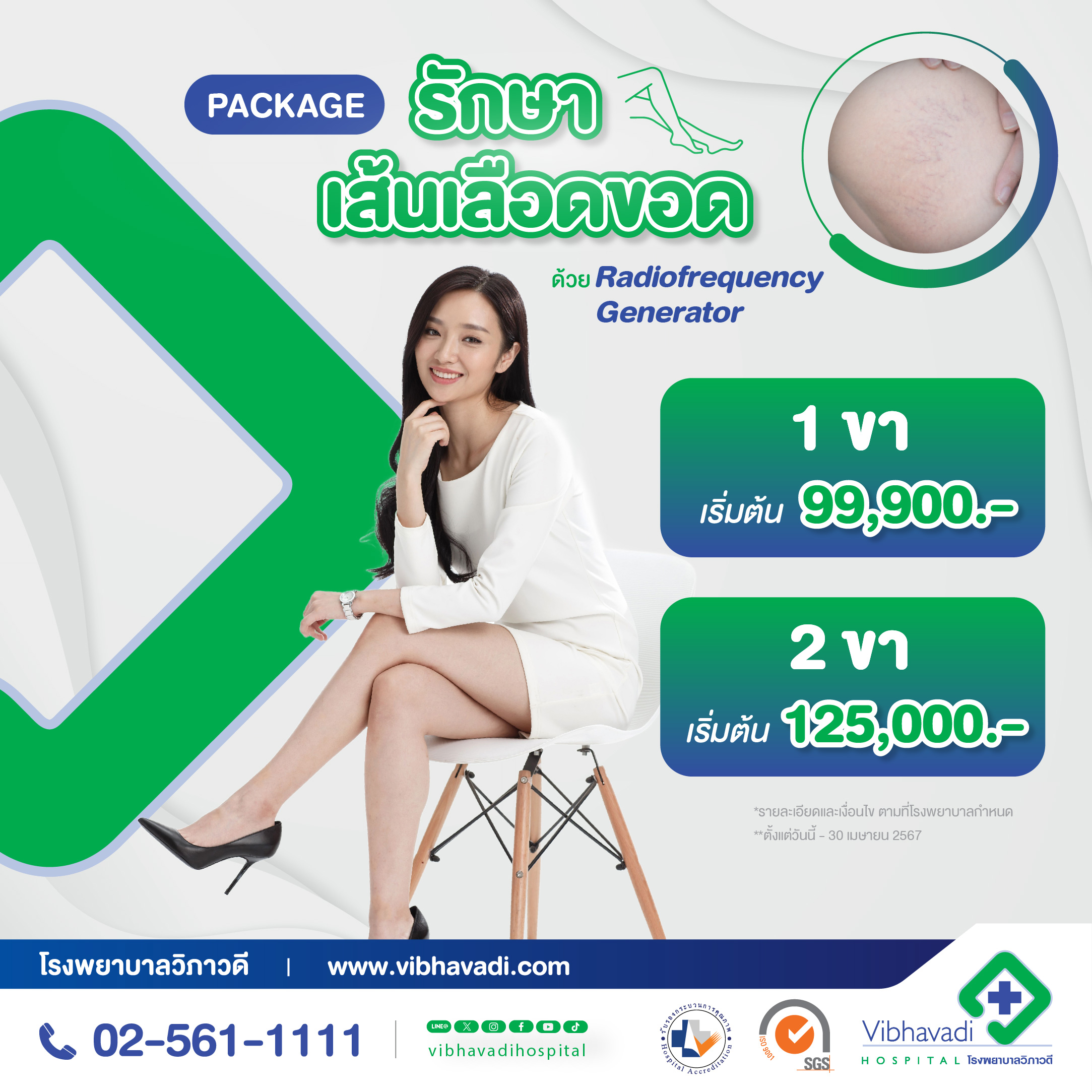
Package for varicose vein surgery using a machine Radiofrequency Generator
Varicose vein surgery using a machine Radiofrequency Generator, which is a modern varicose vein surgery with fast recovery and a small surgical wound.

Package การผ่าตัดข้อกระดูกส่องกล้อง เพื่อรักษาหมอนรองเข่าหรือซ่อมแซม
อาการหมอนรองกระดูกเข่าฉีกขาด สาเหตุเกิดได้หลายอย่าง อาทิ ปวดข้อเข่า เข่าบวม เหยียดงอไม่ได้ ในปัจจุบันการรักษานิยมใช้การผ่าตัดส่องกล้อง Arthroscope แผลผ่าตัดจะมีขนาดเล็ก ลดการเกิดข้อติดยึดในภายหลัง และเพื่อให้ผู้มารับบริการได้ทราบราคาที่ชัดเจน ง่ายกับการตัดสินใจผ่าตัด

Platelet-Rich Plasma (PRP) Injection Treatment for Bone and Knee Problems Package
Bone and Knee Problems Treatment via Platelet-Rich Plasma injection (PRP) doesn’t need to undergo surgery and no joint pain. Treat joint problems such as arthritis, osteoarthritis, or chronic inflammation with PRP. This involves taking plasma enriched with platelets and growth factors that can repair various types of tissues. Approximately 10-2- CC of blood is drawn and spun at an appropriate speed to separate the plasma and platelets from the red blood cells. This resulting solution is then injected into the affected area for natural tissue repair. This treatment aims to reduce pain and accelerate recovery without side effects, residual chemicals, or allergic reactions as the blood used is from the patient’s own body. The effectiveness of the treatment varies depending on the severity and condition of each patient. The decision to use PRP injections will be made according to the examination of the attending physician.

Knee Replacement Surgery Package
Knee replacement surgery is another treatment option. For patients with osteoarthritis who have severe pain Even if treated with medication for a long time or treatment by other means but the symptoms do not improve
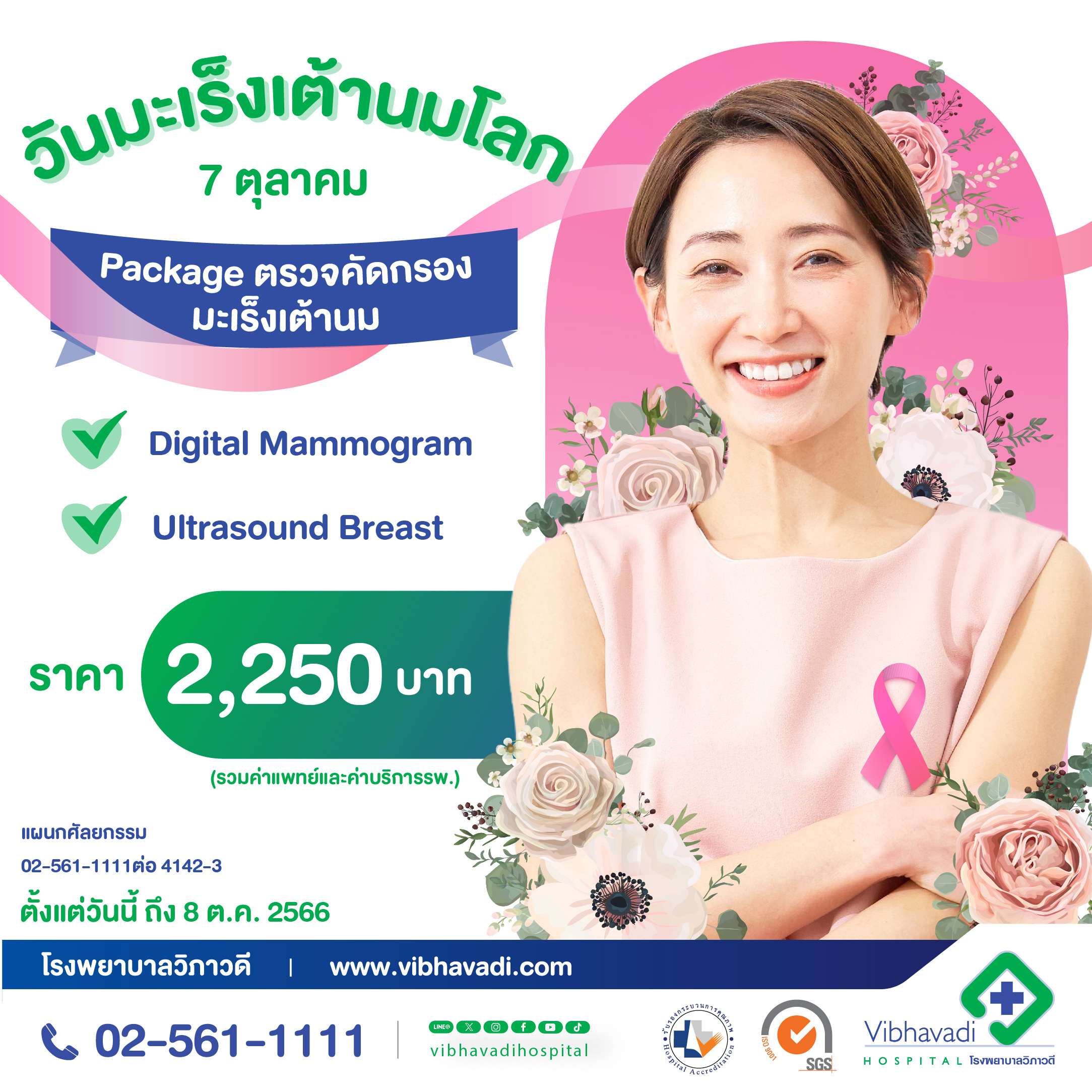
โปรโมชั่น Package ตรวจคัดกรองมะเร็งเต้านม
7 ตุลาคม วันมะเร็งเต้านมสากล (World Breast Cancer Day) มะเร็งเต้านม ภัยเงียบอันดับ 1 ของผู้หญิงทั่วโลก มะเร็งเต้านมป้องกันได้ ด้วยการหมั่นตรวจเต้านมด้วยตนเอง และ พบแพทย์เพื่อตรวจเต้านมอย่างน้อยปีละครั้ง

Package Arthroscopic ACL Knee
Symptoms of a torn knee meniscus can be caused by various factors, such as knee pain, swelling, and inability to fully extend or bend the knee. Currently, the preferred treatment is arthroscopic surgery. The incision from the surgery is small, which reduces the risk of joint stiffness afterwards. To provide clear pricing information and make it easier for patients to decide on surgery, we offer transparent cost details.

Arthroscopic Surgery Package for Frozen Shoulder
Individuals with frozen shoulder experience pain or discomfort when moving their shoulder, with increased pain when attempting to lift their arm. If left untreated for a long time, the muscles around the shoulder can become tight and stiff. Frozen shoulder is often a result of underlying conditions or injuries in the shoulder joint. Therefore, it is important to seek proper examination and treatment.

Package Epidural Steroid Injection (ESI)
Did you know? Injecting steroids into the spinal canal is a treatment that reduces pain from inflammation of the nerves in the spinal canal. This will quickly reduce pain, swelling, and inflammation. without having to perform surgery No need to take painkillers often. This allows patients to return to their normal daily lives faster.

Endoscopic Lumbar Herniated Disc Surgery Package
When you experience tingling sensations similar to being pricked by needles, numbness, or weakness starting from the lower back and radiating through the buttocks down the leg, it may indicate a herniated disc pressing on a nerve. If these symptoms occur, do not leave them untreated for too long. Seek proper examination and treatment to ensure your good health because a good life is one free of illness.

Anterior Cervical Discectomy and Fusion (ACDF) Surgery Package for Cervical Disc Herniation with Nerve Compression
Neck pain is a common condition that can result from various causes. If the pain or numbness extends down to the arm, elbow, or fingers, it may indicate that the cervical spinal nerves are being compressed. Anterior Cervical Discectomy and Fusion (ACDF) surgery may be a suitable option for patients to relieve radiating neck pain down the arm.


