Ultrasound examination
Ultrasound is an examination with high-frequency sound waves by allowing a transducer transmit ultrasound waves to the skin, glands, or tissues with different properties. Those waves will be reflected and scattered back into the transducer (echo). Then, it is recorded, amplified, and adjusted before being sent to the display.
Ultrasound can be used to examine different parts of the body, including:
Used to examine young children under 2 years of age to check the abnormalities in the skull by examining through the fontanelles.
Used to detect abnormalities and find lesions of the thyroid gland, salivary gland, parotid gland, and nodules at the neck area. Moreover, it can be used to examine the carotid artery.
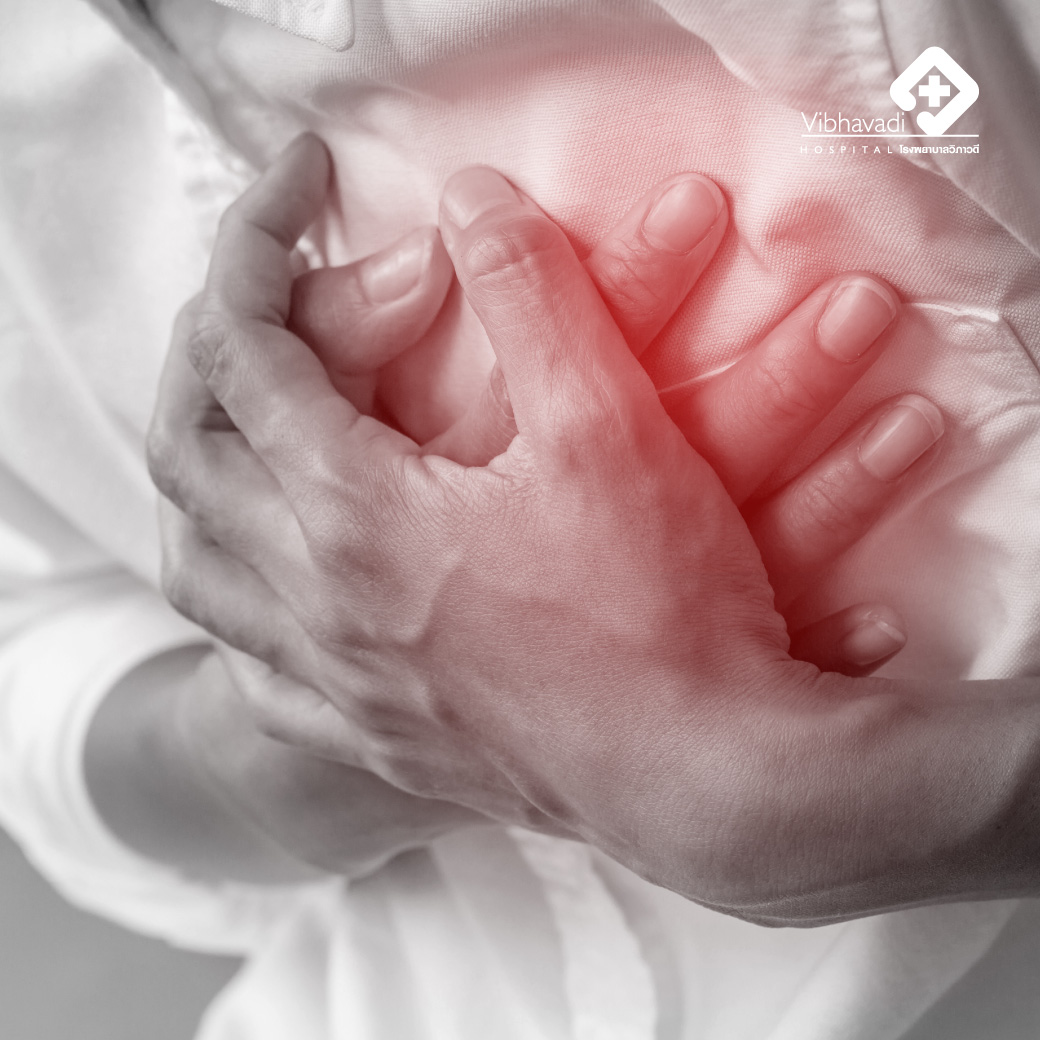
Used to examine for fluid in the pleural space (pleural fluid) or to check for lesions on the chest wall, such as tumors, etc.
Used to examine abnormalities and find lesions of all internal abdominal organs.
Used to investigate abnormalities and lesions in other organs that are soft tissues or have fluid inside, such as muscles. It can also use a Doppler examination of the breast, legs, and large and medium size of blood vessels. Abnormalities of blood vessels, blood flow velocity, vessel blockages are investigated too.
For the head, neck, and thorax, they can be checked immediately without preparation. However, in some children, sedation may be required according to the doctor's orders.
Abdomen
Other parts can be examined immediately. No preparation is required before the examination.
Advice
Privacy Policy | Cookie Policy
Copyright © Vibhavadi Hospital. All right reserved